When
Where
Title
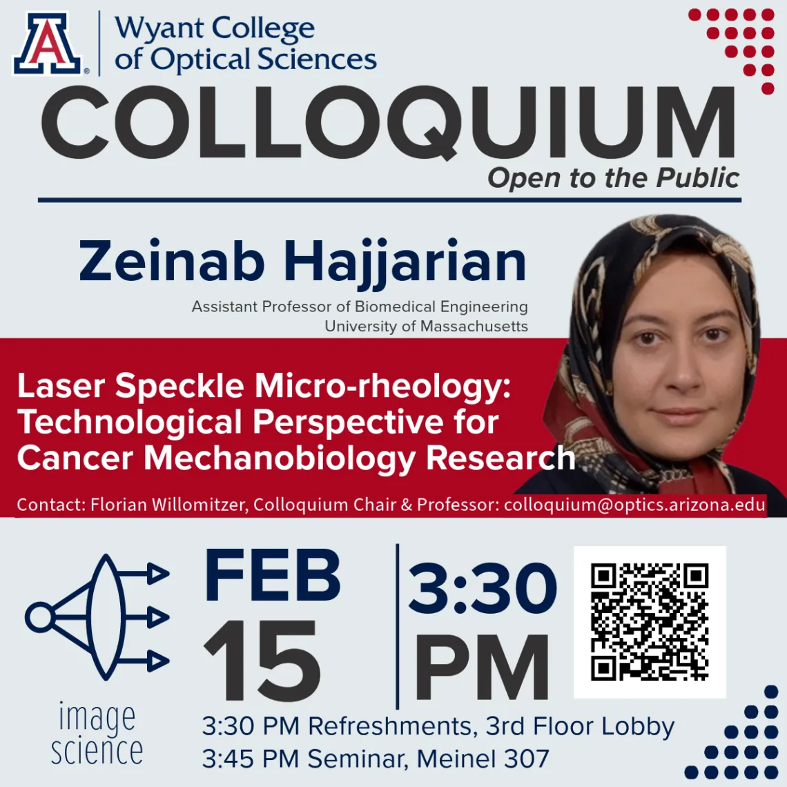
Laser Speckle Micro-rheology: Technological Perspective for Cancer Mechanobiology Research
Abstract
Disease progression in multiple pathological conditions is marked by mechanical alteration of the affected tissue across multiple length scales. For instance, altered mechanical properties of the tumor microenvironment have been raised as both the cause and the consequence of breast carcinogenesis. Increased bulk stiffness of tumors is traditionally exploited to screen for solid tumors in clinical settings via palpation or elastography. However, the tumor microenvironment is not a homogeneous elastic solid. Instead, it exhibits heterogeneous viscoelastic properties that reflect the micro-mechanical interactions of tumor cells and stroma and likely inform the tumor's aggressiveness. Despite this exciting potential, the associations between micro-mechanical features and the clinical prognostic markers of breast carcinoma remain unknown, largely due to the lack of imaging modalities for mapping the micromechanical attributes at length scales perceived by cells. Here, we introduce the concept of laser speckle micro-rheology (LSM). This novel optical approach evaluates the viscoelastic properties of tissues from the intensity fluctuations of the back-scattered light in a non-contact, non-invasive manner. Speckle is a granular intensity pattern formed when a coherent beam of light is scattered from turbid media, such as biological tissue. Speckle fluctuations are exquisitely sensitive to the Brownian motion of the endogenous scattering particles within the tissue and, in turn, the viscoelastic properties of the tissue microenvironment. To prime this technique for applications in cancer mechanobiology research, we have developed the laser Speckle rHEologicAl microscopy (SHEAR), a microscopic embodiment of LSM that transforms the technology from point-sample rheology to a high-resolution imaging modality. The prognostic utility of SHEAR is tested in a large cohort of patient specimens with distinct diagnoses, including a representative number of rare subtypes. Our results demonstrate the links between micromechanical heterogeneities and clinical prognostic markers, including histopathological subtype, tumor grade, receptor expression, and lymph node status in breast carcinoma. These results outline the tremendous potential of SHEAR for both technical maturation and prospective applications in cancer biomechanics and mechanobiology research.
Bio
Zeinab Hajjarian, Ph.D., is an Assistant Professor of Biomedical Engineering at the University of Massachusetts, Lowell. She received her Ph.D. in Electrical Engineering from Pennsylvania State University in 2009. Her doctoral research concentrated on modeling light propagation in the turbid and turbulent atmosphere to enable free-space wireless optical telecommunications. Dr. Hajjarian transitioned to the field of biomedical optics in 2010 when she started her post-doctoral training at the Wellman Center for Photomedicine, Massachusetts General Hospital. Since then, she has focused on developing novel laser speckle micro-rheology (LSM) techniques for rendering and imaging the biomechanical properties of tissues. Dr. Hajjarian has a remarkable record of innovative scholarship. She has published over 20 original manuscripts, with 14 as the first author and two invited review articles. In addition, she has filed more than 15 national and international patent applications, out of which 6 are granted. Her current research interest includes developing novel optical microscopes and sensing devices that permit characterizing the biophysical and biomechanical properties of the soft tissues and biofluids and translating them to the clinical setting for integration in the pipeline of diagnosis.
Can't Join Us In Person?
Register for the Zoom Webinar!
Subscribe to Upcoming Colloquium Announcements
Visit our website for future lecture dates and speaker information
