
Another Wavelength: Travis Sawyer
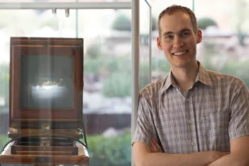
This month in Another Wavelength, we chat with 1st year Ph.D. candidate, Travis Sawyer. Travis also received his B.S. in optical sciences from OSC and is currently mentored by Jennifer Barton.
Where are you from?
I moved around a lot growing up, but consider myself a Tucson native.
What brought you to study optics?
I was always interested in science, particularly astronomy; however, I decided to study optics when I was a freshman at the UA after I received a false-positive diagnosis for leukemia. In the course of that event, I underwent a lot of testing with medical imaging equipment, which kindled my interest in the field of medical imaging. Knowing the reputation of OSC from growing up in Tucson, I thought it would be a good place to build up a foundation to pursue a career in medical imaging research. Since then, my interest has only grown as I continue to learn more and am astounded by the power of light.
Who is your hero in science?
There are a lot of scientists that I look up to and I'm not sure I have a specific hero. I have a tremendous amount of respect for Marie Curie and Lise Meitner, as both of them made profound advances in their field while also facing huge societal barriers. I am also extremely fond of Richard Feynman - not only was he an incredible scientist, but he was a dedicated teacher, public servant, and he also knew how to enjoy life!
Describe your research in 20 words or fewer.
I'm trying to improve early cancer detection by measuring how the optical properties of tissue change throughout disease progression.
Describe your research in 200 words or fewer.
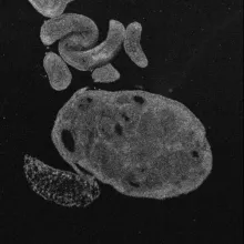
An optical coherence tomography image of an ovary.
My aim is to develop a minimally-invasive method to screen for esophageal cancer. Our current techniques are abysmally ineffective, due in large part to the fact that esophageal cancer doesn't appear to look diseased until it is too late. By measuring different tissue properties using imaging techniques such as hyperspectral imaging (HSI) and optical coherence tomography (OCT), we can get a more complete picture of the tissue health including its structure, chemistry, and metabolism. In particular, hyperspectral imaging gives information regarding how different colors of light interact with the tissue, capturing fluorescence information about the underlying tissue chemistry and also metabolic information such as blood concentration and oxygenation; OCT compliments that data by providing a robust structural reference that we can use to investigate the tissue microstructure. I'm currently working with biopsied samples to characterize the tissue fluorescence so we better understand how the fluorescence emission behaves as the disease evolves. After that, the next step is to design, prototype and test an endoscope (sort of a camera on a flexible arm) that can be inserted into the esophagus to image the tissue using HSI and OCT.
Name three neat facts about you.
- I started my own company as an undergraduate.
- I have lived in Stuttgart, Germany; Amsterdam, Netherlands; and Cambridge, England.
- I helped authenticate several paintings including a Van Gogh using infrared and X-ray light to image underneath the layers of paint and determine the artwork's origin and history.