
Date Published: November 24, 2014
Optical imaging techniques are now frequently employed for medical diagnosis. A familiar example is biopsy, where a pathologist observes an excised tissue sample under an optical microscope to diagnose a disease. The discipline of optical biopsy takes an optical instrument (e.g., a confocal microscope) in a miniaturized endoscopic form directly to the tissue in need of evaluation. Optical biopsy allows real-time in situ analysis with greater ease and, potentially, increased accuracy. Arthur Gmitro’s research group has pioneered the development of fiber-bundle-based fluorescence confocal microendoscopy, building systems to image a variety of endoscopically and/or laparoscopically accessible organ sites. The instrument shown in the figure at above left is being clinically evaluated for its ability to identify early-stage ovarian and fallopian tube cancers.
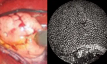
Left: Confocal microendoscope imaging probe in contact with an ovary surface during a laparoscopic procedure. Right: Fluorescence image obtained from the probe showing individual nuclei of cells on the epithelial ovary surface.