Date Published: August 28, 2024
Researchers, led by Judith Su, associate professor of optical sciences and biomedical engineering; have developed a groundbreaking biological sensing method capable of detecting substances at zeptomolar levels, an incredibly miniscule scale. The method, using Su's FLOWER device (Frequency Locked Optical Whispering Evanescent Resonator), is label-free, meaning it does not require tagging substances with fluorescent or radioactive markers.
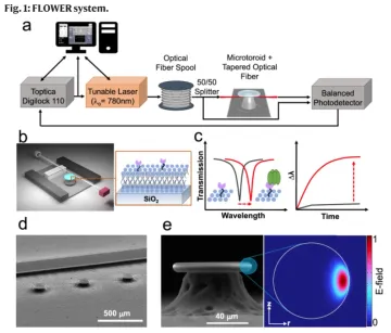
FLOWER utilizes a microtoroid optical resonator, which captures target compounds and detects changes in light wavelength to sense ultra-low concentrations. In experiments, the researchers focused on the kappa-opioid receptor, which plays a key role in pain management. FLOWER’s sensitivity could lead to the discovery of more effective drugs that may be overlooked with current technologies. The research was published in Nature Communications and is supported by grants from the NIH.
Experts believe this represents a major leap forward in biosensing, with potential applications in drug discovery, toxin detection, and cancer screening. Su and her collaborators at UCLA won a $650,000 Phase 1, National Science Foundation Convergence Accelerator grant last year to continue their investigations. Convergence Accelerator grants are awarded specifically to speed scientific research likely to produce beneficial applications for society.

a A tunable laser is coupled to the microtoroid cavity through a tapered optical fiber. b Schematic diagram of the constructed biosensing chamber (top cover glass not shown). The tapered fiber is glued to the support wall with a polymer adhesive. Inset shows the structure of a membrane protein (pink) incorporated in a lipid bilayer (blue) on the toroid’s glass surface. c Sketches of how the resonant wavelength red-shifts when analytes adsorb onto the functionalized toroid surface (inset). d SEM image of microtoroid array alongside a support wall. The scale bar is 500 µm. e SEM side view image of a microtoroid structure. The scale bar is 40 µm. The inset picture shows a COMSOL simulation of an optical mode of the lipid coated cavity in a buffered aqueous solution.
Adley Gin
